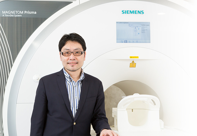
Team Director
Takuya Hayashi
M.D., Ph.D.
Laboratory for Brain Connectomics Imaging
Location Kobe / MI R&D Center
E-mailtakuya.hayashi@riken.jp

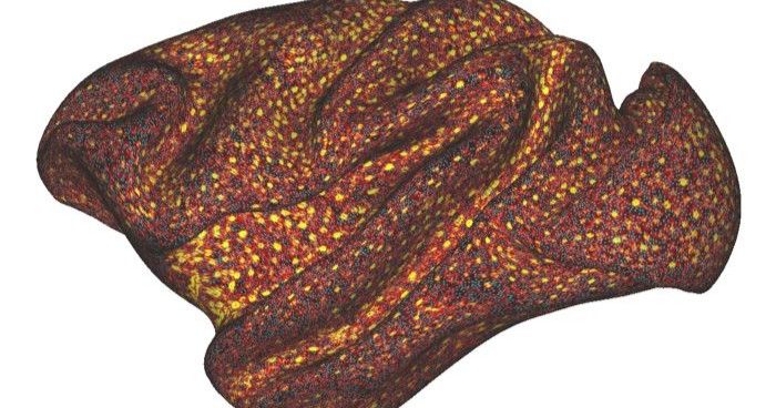
Our laboratory develops neuroimaging research techniques using magnetic resonance imaging (MRI) to comprehensively understand organization of brain function, structure, connectivity (brain connectome) in human and non-human primates. By visualizing the living brains, we investigate how development, aging, evolution, social behaviors, genetics, environments, dysfunction, and plasticity are represented in the dynamics and reorganization of the brain system. Strength of noninvasive imaging methods is that it allows us to observe brain systems across individuals, time and species, which may provide us a clue to understand how individual variability is formed, how we develop and spend our life, and how brains have evolved across primate species. However, quality of noninvasive imaging methods including MRI is often hampered by its limited resolution and addressing neurobiological findings is easily contaminated by various artifacts originated from living organs. Observed findings rarely have direct link to those found in traditional neuroscientific methods (microscopy, electrophysiology) – electromagnetic phenomenon visualized by MRI must be translated into those of neurobiological events.
Our lab has developeda high-resolution imaging system to visualize functional anatomy of non-human primate brains.This was achieved by combination of various technical developments including high-resolution signal detection, accelerated imaging, modelling, noise reduction and accurate mapping. Our development achieved the highly sensitive and reliable estimation of functional connectivity as compared with any other research facilities, thereby we advance the international research project for non-human primate neuroimaging and neuroanatomy (NHP_NNP). This project aims to harmonize the MRI technologies between NHP and humans, and to understand the general principle of the primate brain organization and pathophysiology in the primate animal models of human brain diseases. We make use of our developments in NHP for human brain imaging for understanding brain system and diseases, which is supported by a Brain/MINDS-beyond project. Our research and technology are based on close domestic and international collaborations. Please do not hesitate to contact us if you would work together with us to develop brain MRI techniques and understand the brain functional anatomy, plasticity, and pathology.
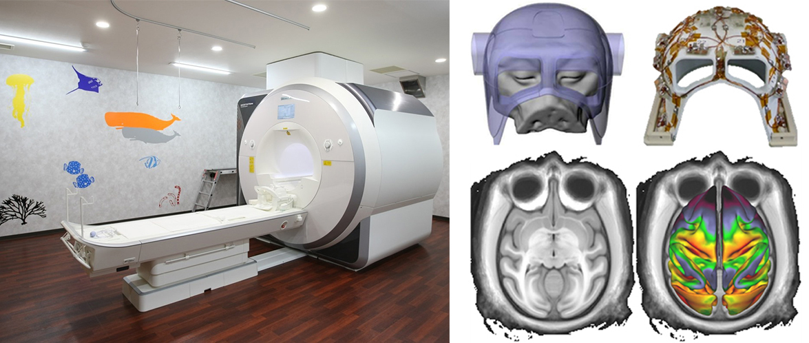
Primate Connectome MRI scanner installed in RIKEN Kobe (left) and our developed radiofrequency receiving coil and brain/head images of non-human primates (right)
Our lab for brain connectomicsimaging aims to visualize brain structure, function and connectivity using MRI and to comprehensive understanding of dynamics of brains in human and non-human primates
Research Theme
- Visualization of brain connectomics
- Reorganization of brain network in regeneration
- Reorganization of brain network in plasticity
- Sociality, brain network and neurotransmitter
Selected Publications
Autio JA, Kimura I, Ose T, et al.
Mapping vascular network architecture in primate brain using ferumoxytol-weighted laminar MRI.
eLife (in press)
(2024)
Ikeda T, Autio JA, Kawasaki A, et al.
Cortical adaptation of the night monkey to a nocturnal niche environment: a comparative non-invasive T1w/T2w myelin study.
Brain Structure & Function
228(5), 1107-1123 (2023)
doi: 10.1007/s00429-022-02591-x
Ose T, Autio JA, Ohno M, et al.
Anatomical variability, multi-modal coordinate systems, and precision targeting in the marmoset brain.
Neuroimage
250, 118965 (2022)
doi: 10.1016/j.neuroimage.2022.118965
Hayashi T, Hou Y, Glasser MF, et al.
The nonhuman primate neuroimaging and neuroanatomy project.
NeuroImage
229, 117726 (2021)
doi: 10.1016/j.neuroimage.2021.117726
Autio JA, Glasser MF, Ose T, et al.
Towards HCP-Style macaque connectomes: 24-Channel 3T multi-array coil, MRI sequences and preprocessing.
NeuroImage
215, 116800 (2020)
doi: 10.1016/j.neuroimage.2020.116800
Aso T, Sugihara G, Murai T, et al.
A venous mechanism of ventriculomegaly shared between traumatic brain injury and normal ageing.
Brain : a journal of neurology
143(6), 1843-1856 (2020)
doi: 10.1093/brain/awaa125
Fukutomi H, Glasser MF, Zhang H, et al.
Neurite imaging reveals microstructural variations in human cerebral cortical gray matter.
NeuroImage
182, 488-499 (2018)
doi: 10.1016/j.neuroimage.2018.02.017
Morizane A, Kikuchi T, Hayashi T, et al.
MHC matching improves engraftment of iPSC-derived neurons in non-human primates.
Nature Communication
8, 385 (2017)
doi: 10.1038/s41467-017-00926-5
Kikuchi T, Morizane A, Doi D, et al.
Human iPS cell-derived dopaminergic neurons function in a primate Parkinson’s disease model.
Nature
548, 592-596 (2017)
doi: 10.1038/nature23664
Takenobu Y, Hayashi T, Moriwaki H, et al.
Motor recovery and microstructural change in rubro-spinal tract in subcortical stroke.
NeuroImage: Clinical
14, 201-208 (2014)
doi: 10.1016/j.nicl.2013.12.003
Yokoyama C, Kawasaki A, Hayashi T, et al.
Linkage Between the Midline Cortical Serotonergic System and Social Behavior Traits: Positron Emission Tomography Studies of Common Marmosets.
Cerebral Cortex
23(9), 2136-2145 (2013)
doi: 10.1093/cercor/bhs196
Hayashi T, Ko JH, Strafella AP, et al.
Dorsolateral prefrontal and orbitofrontal cortex interactions during self-control of cigarette craving.
Proceedings of the National Academy of Sciences of the United States of America
110(11), 4422-4427 (2013)
doi: 10.1073/pnas.1212185110
Hayashi T, Wakao S, Kitada M, et al.
Autologous mesenchymal stem cell-derived dopaminergic neurons function in parkinsonian macaques.
Journal of Clinical Investigation
123(1), 272-284 (2013)
doi: 10.1172/JCI62516
Hayashi T, Shimazawa M, Watabe H, et al.
Kinetics of neurodegeneration based on a risk-related biomarker in animal model of glaucoma.
Molecular neurodegeneration
8, 4 (2013)
doi: 10.1186/1750-1326-8-4
Ikoma Y, Watabe H, Hayashi T, et al.
Measurement of density and affinity for dopamine D(2) receptors by a single positron emission tomography scan with multiple injections of [(11)C]raclopride.
Journal of cerebral blood flow and metabolism : official journal of the International Society of Cerebral Blood Flow and Metabolism
30(3), 663-73 (2010)
doi: 10.1038/jcbfm.2009.239
Takagi Y, Takahashi J, Saiki H, et al.
Dopaminergic neurons generated from monkey embryonic stem cells function in a Parkinson primate model.
Journal of Clinical Investigation
115(1), 102-109 (2005)
doi: 10.1172/JCI21137
Members
Takuya Hayashi
Team Director
Toshihiko Aso
Deputy Team Director
Takuro Ikeda
Research Scientist
Akiko Uematsu
Research Scientist
Naofumi Yoshida
Research Associate
Takayuki Ose
Technical Scientist
Takashi Azuma
Technical Scientist
Masahiro Ohno
Expert Technician
Tomoko Ishibuchi
Technical Staff I
Reiko Kobayashi
Technical Staff I
Tomoko Oyanagi
Assistant
Natsuko Kashida
Research Part-time Worker I
Rei Hashimoto
Student Trainee
Arihito Nauchi
Student Trainee